Andrological
It is the medical specialty that deals with male health, particularly relating to the problems of the male reproductive system problems.
The male has reproductive organs, they include: testes, ducts epididymis and vas deferens, accessory gland seminal vesicles, prostate On reaching sexual maturity, testes make and store millions of tiny sperm cells. They also produce testosterone.
Testosterone is the hormone that causes boys to develop deeper voices, bigger muscles, and body and facial hair. It also stimulates the production of sperm.

Epididymis and the vas deferens transport sperm. The epididymis and the testicles hang in a pouch-like structure outside the pelvis called the scrotum. This bag of skin helps to regulate the temperature of testicles, which need to be kept cooler than body temperature to produce sperm.
The accessory glands, including the seminal vesicles and the prostate gland, provide fluids that lubricate the duct system and nourish the sperm. The urethra is the channel that carries the sperm (in fluid called semen) to the outside of the body through the penis.
The penis is actually made up of two parts: the shaft and the glans. The shaft is the main part of the penis and the glans is the tip . At the end of the glans is a small slit or opening, which is where semen and urine exit the body through the urethra. The inside of the penis is made of a spongy tissue that can expand and contract.
Andrological issues in the pregnant patient present a unique clinical dilemma. These patients may be challenging to treat due to risks associated with medications and surgical procedures. This review aims to provide an update on the physiological changes and surgical risks in pregnancy. In addition, we review the approach for management of Andrological and urinary tract infections in pregnancy. Lastly, we highlight the importance of a multidisciplinary approach to placenta percreta, a condition not commonly addressed in Andrological education.
Spermatogenesis
Spermatogenesis is the production of sperm from the primordial germ cells. Once the vertebrate Premordial Germ Cells arrive at the genital ridge of a male embryo, they become incorporated into the sex cords. They remain there until maturity, at which time the sex cords hollow out to form the seminiferous tubules, and the epithelium of the tubules differentiates into the Sertoli cells. The initiation of spermatogenesis during puberty is probably regulated by the spermatogenic germ cells, the spermatogonia. The germ cells begin to differentiate.The spermatogenic germ cells are bound to the Sertoli cells by N-cadherin molecules on both cell surfaces and by galactosyltransferase molecules on the spermatogenic cells that bind a carbohydrate receptor on the Sertoli cells. The Sertoli cells nourish and protect the developing sperm cells, and spermatogenesis.
After reaching the gonad, the PGCs divide to form type A1 spermatogonia. The A1 spermatogonia are found adjacent to the outer basement membrane of the sex cords. Thus, each type A1 spermatogonium is a stem cell capable of regenerating itself as well as producing a new cell type. The A2 spermatogonia divide to produce the A3 spermatogonia, which then beget the type A4 spermatogonia. It is possible that each of the type A spermatogonia are stem cells, capable of self-renewal. The A4 spermatogonium has three options: it can form another A4 spermatogonium (self-renewal); it can undergo cell death (apoptosis); or it can differentiate into the first committed stem cell type, the intermediate spermatogonium. Intermediate spermatogonia are committed to becoming spermatozoa, and they divide mitotically once to form the type B spermatogonia. These cells are the precursors of the spermatocytes and are the last cells of the line that undergo mitosis. They divide once to generate the primary spermatocytes—the cells that enter meiosis.
Each primary spermatocyte undergoes the first meiotic division to yield a pair of secondary spermatocytes, which complete the second division of meiosis. The haploid cells thus formed are called spermatids. During the divisions from type A1 spermatogonium to spermatid, the cells move farther and farther away from the basement membrane of the seminiferous tubule and closer to its lumen. Each type of cell can be found in a particular layer of the tubule. The spermatids are located at the border of the lumen, and here they lose their cytoplasmic connections and differentiate into sperm cells. In humans, the progression from spermatogonial stem cell to mature sperm takes 65 days.
Spermiogenesis
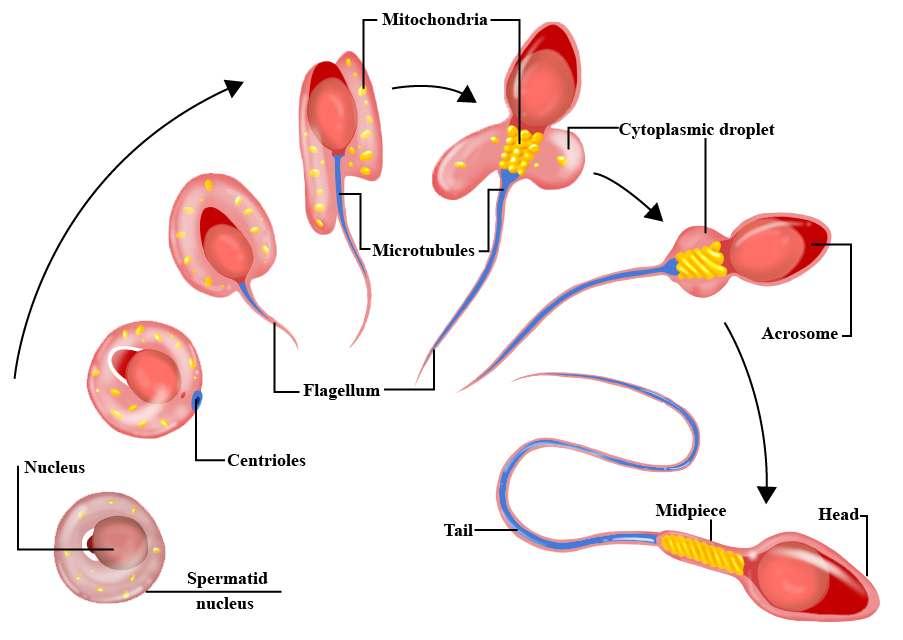
One of the major changes in the nucleus is the replacement of the histones by protamines. During spermiogenesis, the nucleosomes dissociate, and the histones of the haploid nucleus are eventually replaced by protamines. This causes the complete shutdown of transcription in the nucleus and facilitates its assuming an almost crystalline structure. The resulting sperm then enter the lumen of the tubule.
Each day, some 100 million sperm are made in each human testicle, and each ejaculation releases 200 million sperm.
Defects in spermiogenesis and spermiation are manifested as low sperm number, abnormal sperm morphology and poor motility.
Azoospermia
It means no sperm in the ejaculate. Around 10 percent of infertile men and 1 percent of all men have azoospermia. It may be obstructive or non obstructive i.e. blockage preventing sperms to reach ejaculate or decreased sperm production by testes.There may be many potential causes, including some genetic conditions such as Klinefelter’s syndrome, medical treatments such as chemotherapy or radiation, recreational drugs such as some narcotics, and anatomical abnormalities such as varicoceles or absence of the vas deferens on each side. There may be surgical causes like a vasectomy, which prevents sperm from joining other fluids in the ejaculate. In most cases, though, azoospermia is likely due to factors such as genetic conditions, poor testicular development or environmental toxins.
Azoospermia Treatment
The best treatment is a customized approach based on many factors, such as partner’s age and reproductive function, physical exam findings, blood test results, long- and short-term family goals and even finances. Depending on the suspected causes, many treatments may be available. If there is a blockage (or history of vasectomy), reconstruction might be the best treatment for some men. In others, removing offending agents such as medications or recreational drugs might be the first step. Sometimes hormonal abnormalities may need correction. In some men, treatment will increase sperm production. In some others, surgery to fix anatomical abnormalities or varicoceles can be pursued, and in others the best option is to go directly into the testicle to attempt retrieval of sperm that could be used for ART. Finally, some couples may require donor sperm or adopting an infant or child.PESA
 Percutaneous Epididymal Sperm Aspiration is a procedure performed for men who are having sperm retrieved for IVF/ICSI who have obstructive azoospermia from either a prior vasectomy or infection. It is done with local anesthesia in the operating room or office and is coordinated with their female partner’s egg retrieval. Sperms are aspirated from epididymis.